- About Us
Fact SheetOverseas Offices & RepresentativesAccreditations and AwardsPatient Stories
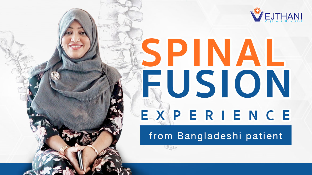
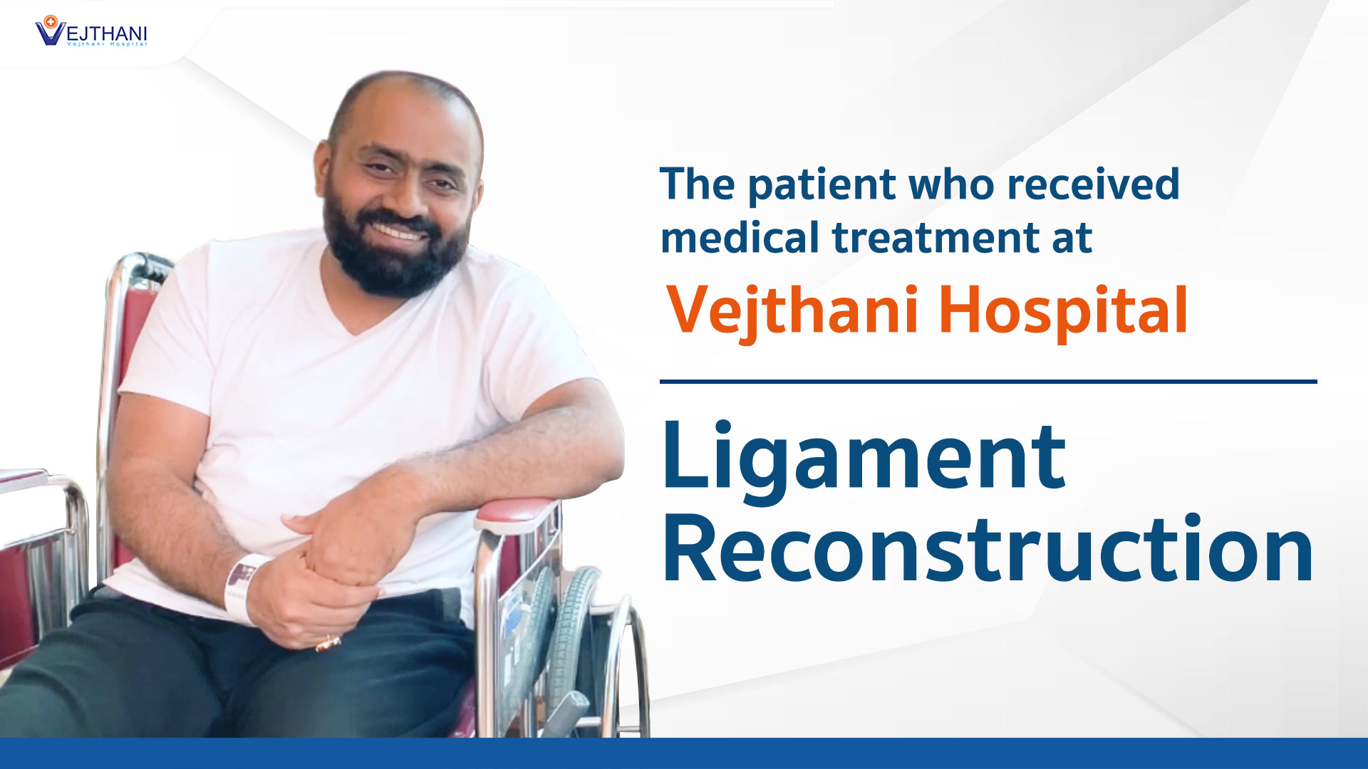
 Spinal fusion experience from Bangladeshi patientMyanmar Actress Patricia’s Intraocular Lens Surgery Experience at Vejthani HospitalLigament ReconstructionA comprehensive kid’s checkup at Vejthani Hospital’s Super Kid’s Center
 - Patient Services
- Medical Departments & Centers
- Packages & Programs
- Health Information
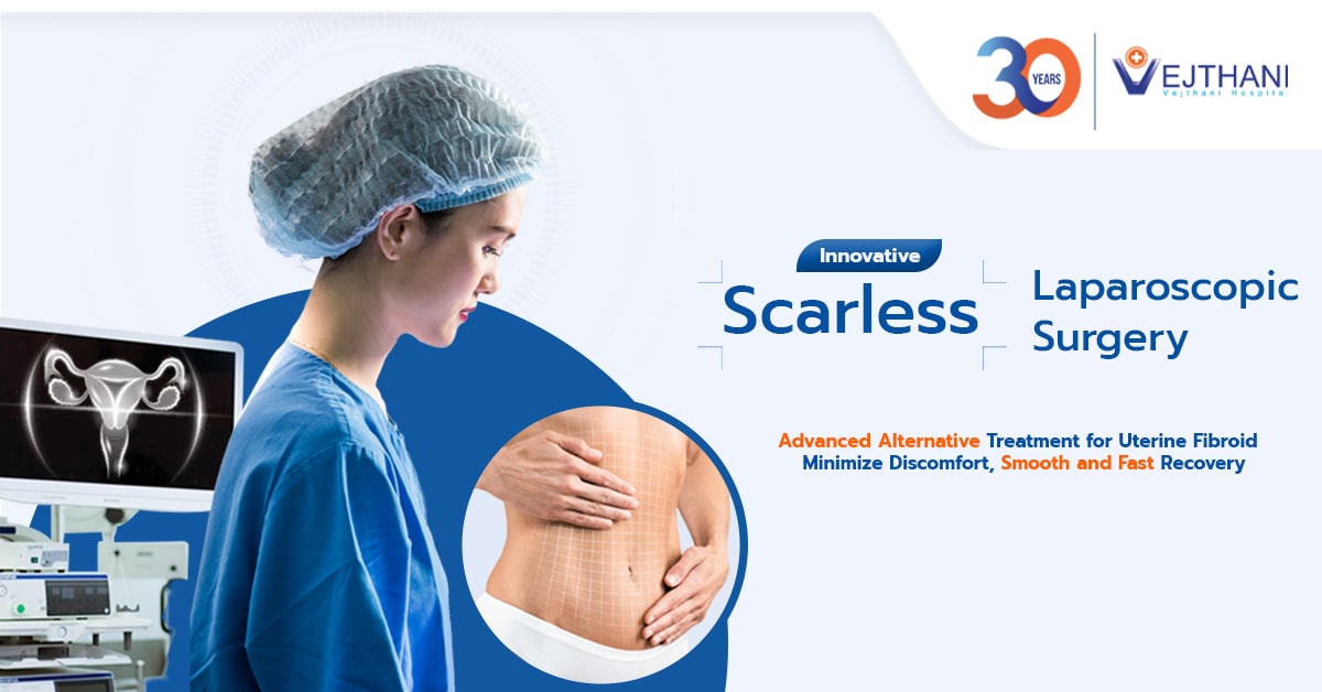
Health ArticlesHealth VideosNews and UpdatesSummer’s Silent Killer: Your Essential Guide to Preventing and Defeating HeatstrokeGroundbreaking Approach to Treat Cerebral Aneurysm: Biplane DSA and the Future of NeurointerventionScarless Laparoscopic Surgery: Advanced Treatment for Uterine FibroidTotal Knee Replacement with Technological Breakthrough – Rosa Robotic-Assisted Surgery


- About Us
Fact SheetOverseas Offices & RepresentativesAccreditations and AwardsPatient StoriesSpinal fusion experience from Bangladeshi patientMyanmar Actress Patricia’s Intraocular Lens Surgery Experience at Vejthani HospitalLigament ReconstructionA comprehensive kid’s checkup at Vejthani Hospital’s Super Kid’s Center
- Patient Services
- Medical Departments & Centers
- Packages & Programs
- Health Information
Health ArticlesHealth VideosNews and UpdatesSummer’s Silent Killer: Your Essential Guide to Preventing and Defeating HeatstrokeGroundbreaking Approach to Treat Cerebral Aneurysm: Biplane DSA and the Future of NeurointerventionScarless Laparoscopic Surgery: Advanced Treatment for Uterine FibroidTotal Knee Replacement with Technological Breakthrough – Rosa Robotic-Assisted Surgery
Neuroscience Center
Vejthani Hospital’s Neuroscience Center is an integrated center that provides neurological services comprising treatment of all neurological conditions, including migraines, headaches and Parkinson’s disease, as well as brain tumor removal and pain management.
This is a major achievement of our medical services in which we have many experienced and qualified neurologists and neurosurgeons of different sub-specialties.
Our highly-qualified team includes experienced neurologists and neurosurgeons with many different sub-specialties. This allows us to address a range of brain-related health concerns and provide the best medical care for our patients.
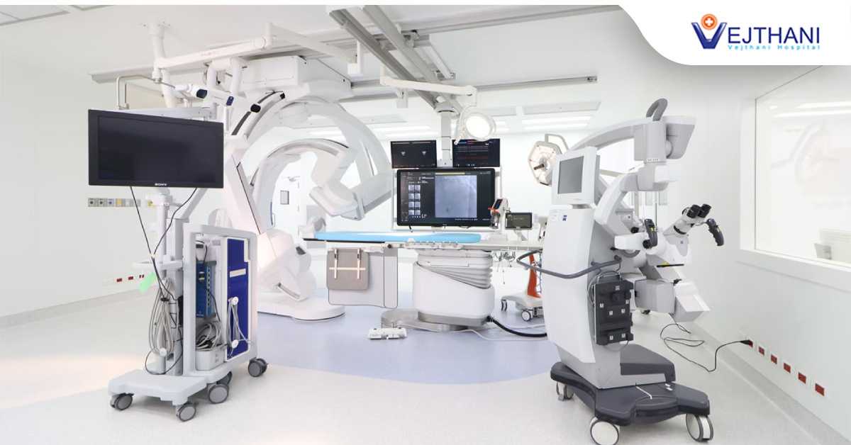
Vejthani Hospital’s Neuroscience Center offers both out-patient and in-patient services to meet the specific needs of our patients. We have state-of-the-art medical equipment so we can provide top-quality care using the most modern methods.
Patients can be assured of receiving an excellent international level of care for both selective and emergency needs. Our dedicated and experienced staff, up-to-date technologies and modern facilities make Vejthani Hospital the best place for brain tumor treatment in Thailand.
SERVICE HOURS
Open Daily: 08.00 am – 05.00 pm
LOCATION
Neuroscience Center, 4th Floor, King of Bones Building, Vejthani Hospital
APPOINTMENTS & INQUIRIES
Call: +66 (0) 2734-0000 ext. 5400
Services
Our services can be divided into three main areas:
1) Neurology:
This branch of medicine deals with treating disorders of the nervous systems, such as:
- Ischemic stroke (caused by lack of blood flow)
- Hemorrhagic stroke (caused by bleeding)
- Thrombotic stroke
- Emergency stroke
- Headaches and facial pain
- Vertigo and syncope
- Chronic pain
- Sleep disorders
- Movement disorders, including Parkinson’s disease
- Dementia and behavioral disorders
- Diabetic neuropathy
- Epilepsy
2) Neurosurgery
This branch specializes in the diagnosis and surgical treatment of disorders of the central and peripheral nervous system, such as:
- Brain and spinal cord tumors
- Brain and spinal cord injuries
- Interventional pain management and neuromodulation
3) Neurodiagnostics
This branch offers diagnostic services to people of all ages with a nervous system disorder. Neurophysiologic studies include:
- Electroencephalography (EEG), which covers both routine and 24-hour video EEG monitoring
- Polysomnography (PSG) or sleep lab studies
- Electromyogram (EMG) or nerve conduction studies (NCS)
- Neurosonology, including carotid doppler and transcranial doppler (TCD)
- Neuroradiology, with techniques such as:
- CT scanning which provides more detailed information on head injuries, strokes, brain tumors and other brain diseases.
- Advanced 3D magnetic resonance imaging, which comes with acoustic reduction technology to drastically reduce humming sounds and make examinations easier, and other advanced technologies to provide high-definition images of the brain that are necessary for treatment.
- Magnetic resonance imaging (MRI) of the brain, which uses a powerful magnetic field, radio waves and a computer to produce detailed pictures of the brain and other cranial structures that are clearer and more detailed than other imaging methods.
- Magnetic resonance angiogram (MRA), a type of magnetic resonance imaging (MRI) scan that uses a magnetic field and pulses of radio wave energy to provide pictures of blood vessels inside the brain.
- Selective cerebral angiography is a diagnostic test that uses x-rays and an iodine-containing contrast material to produce pictures of blood vessels in the brain selectively via catheter.
- Computerized transaxial angiography (CTA), a less invasive diagnostic test that uses intravenous contrast injection to identify blood vessels in the brain and neck. This test can be done within half an hour.